


A 57-year-old male presented with bilateral bradykinesia, resting tremor, more pronounced in his right hand, and episodes of freezing upon initiating walking. During the visit, the family reported the patient having speaking, crying and moving his arms and legs at night, as well as experiencing excessive daytime sleepiness (EDS).
Symptoms appeared 4 years ago, initially with rigidity, bradykinesia and gait disturbances, followed 2 years later by sleep disturbances. The patient’s motor function progressively deteriorated, and memory problems and occasional visual hallucinations (VH) were also reported. During the visit, we assessed the patient’s cognitive status using the Montreal Cognitive Assessment (MoCA), obtaining a score of 18 out of 30 (dementia) [1]. His brain magnetic resonance imaging (3.0T MRI) revealed cerebral atrophy with ex vacuo hydrocephalus and small gliotic changes.
Upon initial presentation, the patient had Parkinsonism with resting tremor, axial and limb rigidity, right side predominance, as well as gait disturbance with start hesitation and freezing episodes, mainly on turning. At that time, the diagnosis was unclear with most likely ones being Parkinson’s disease dementia (PDD) or dementia with Lewy bodies (DLB). Antiparkinsonian treatment started with an initial dose of levodopa/carbidopa at 375 mg/day and donepezil at 5 mg/day.
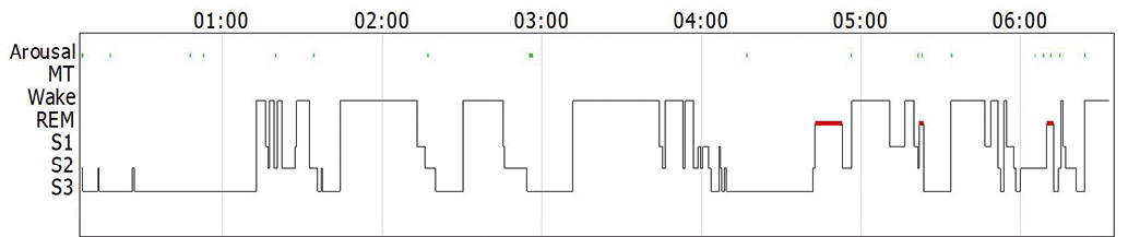
Given the patient’s poor sleep quality due to nighttime behavioral disturbance, rapid eye movement (REM) sleep behavior disorder (RBD) was suspected. He underwent polysomnography (PSG), but surprisingly, REM sleep without atonia (RSWA), a PSG marker of RBD, was absent. All behavioral episodes occurred during wakefulness, with the patient experiencing disturbing VH accompanied by speech, crying, and movements. Mild obstructive sleep apnea (apnea-hypopnea index 8.3/h) was also diagnosed. Polysomnography revealed no episodes of REM without atonia or RBD, probably due to the low proportion of REM sleep (5.7%) and prolonged periods of wakefulness (Fig. 1).
During the follow-up visit, quetiapine 25mg at bedtime was added to the treatment, which significantly improved the patient’s sleep quality and partially increased his daytime alertness.
The patient’s MoCA score decreased from 18 to 8 out of 30 after one year after the initial examination. His Parkinsonism symptoms progressed, requiring an increase in the levodopa dose.
The patient was diagnosed with probable DLB according to the revised diagnostic criteria for DLB [2]: dementia and core criteria including recurrent VH and Parkinsonism in the absence of additional biomarkers (Table 1).
DISCUSSION
Nocturnal behavioral activity can have many etiologies, such as RBD, other parasomnias, nighttime confusion, and VH. Some of these can co-occur in the same patient, especially in those with cognitive impairment.
Rapid eye movement sleep behavior disorder is a REM sleep parasomnia in which the muscle atonia normally present during REM sleep is absent, resulting in the patient “acting out their dreams,” exhibiting a variety of verbal and motor activities [2]. Rapid eye movement sleep behavior disorder affects less than 1% of the adult population but can be found in up to 25-50% of patients with synucleinopathies such as Parkinson’s disease, DLB and multiple system atrophy. It often precedes Parkinsonian motor signs by several years and is considered as a prodromal biomarker of these disorders [3]. About 80% of patients diagnosed with DLB have RBD, and in majority of them it appears before the onset of dementia and Parkinsonism [4]. Polysomnographically confirmed RBD has been included in the DLB diagnostic criteria as an indicative biomarker [3]. Unlike most cases, we did not find RBD or REM without atonia in our patient during PSG as a supporting feature, possibly due to lack of REM sleep and prolonged periods of wakefulness seen on the hypnogram (Fig. 1). The latter could be attributed to the so-called “first-night” effect, when the polysomnogram is affected by the fact of sleeping in a new environment with additional disturbing factors (wires, electrodes, etc.).
Visual hallucinations are most common in patients with PDD and DLB, although hallucinations of other modalities, such as acoustic, tactile, and olfactory can also occur. Visual hallucinations can vary in their phenomenology, quality, frequency, insight, time of day, and emotional impact on the patient [5]. Patients with PDD or DLB often see various objects, children, miniature or normal-size people, etc., or experience a sensation of someone’s presence or passing by [5]. In some cases, nighttime hallucinations can lead to a state of confusion where patients interact with VH and may become agitated [5,6]. Although in some cases this can be attributed to RBD, in our patient it occurred in a waking state, recorded during PSG.
We diagnosed the patient with DLB based on the presence of dementia, cognitive fluctuations, VH, and Parkinsonism. These symptoms met the revised clinical diagnostic criteria for probable DLB [2].
A prerequisite for the diagnosis of DLB is dementia, defined as a progressive cognitive decline of sufficient magnitude to interfere with the performance of normal social or occupational functions.
Our case report has some limitations. The patient had short REM sleep during the recording night, which may have prevent us from detecting RSWA and RBD-related behaviors, which could have been further improved with additional nights of recording.
Differentiation between DLB and PDD is debatable because of many overlaps between these disorders and the focus on the chronology dementia and Parkinsonism [7]. As demonstrated in this clinical case, it can also be hard to diagnose RBD in these patients. While PDD or DLB patients with better cognitive function can provide necessary details of VH, this may not be true for patients with significant dementia [8]. Being a gold standard of sleep examination, PSG can help in identifying nighttime disorder and improve treatment for these complex patients. In our patient’s case, a low dose of an atypical antipsychotic before bedtime resolved his nocturnal behavioral activity, eventually improving his daytime alertness.
Table 1. Revised criteria for the clinical diagnosis of probable and possible dementia with Lewy bodies [2].
Essential criteria: dementia, defined as a progressive cognitive decline that is severe enough to interfere with normal social or occupational functions, or with usual daily activities. Prominent or persistent memory impairment may not necessarily occur in the early stages but is usually evident with progression. Deficits on tests of attention, executive function, and visuoperceptual ability may be especially prominent and occur early. |
Core clinical features (The first 3 typically occur early and may persist throughout the course.)
|
Supportive clinical features Severe sensitivity to antipsychotic agents; postural instability; repeated falls; syncope or other transient episodes of unresponsiveness; severe autonomic dysfunction, e.g., constipation, orthostatic hypotension, urinary incontinence; hypersomnia; hyposmia; hallucinations in other modalities; systematized delusions; apathy, anxiety, and depression. |
Indicative biomarkers
|
Supportive biomarkers
|
Probable dementia with Lewy bodies (DLB) can be diagnosed if: a. Two or more core clinical features of DLB are present, with or without the presence of indicative biomarkers, or b. Only one core clinical feature is present, but with one or more indicative biomarkers. Probable DLB should not be diagnosed on the basis of biomarkers alone. |
Possible DLB can be diagnosed if: a. Only one core clinical feature of DLB is present, with no indicative biomarker evidence, or b. One or more indicative biomarkers are present but there are no core clinical features. |
Dementia with Lewy bodies is less likely: a. In the presence of any other physical illness or brain disorder including cerebrovascular disease, sufficient to account in part or in total for the clinical picture, although these do not exclude a DLB diagnosis and may serve to indicate mixed or multiple pathologies contributing to the clinical presentation, or b. If Parkinsonian features are the only core clinical feature and appear at a first time at a stage of severe dementia. Dementia with Lewy bodies should be diagnosed when dementia occurs before or concurrently with Parkinsonism. The term Parkinson disease dementia (PDD) should be used to describe dementia that occurs in the context of well-established Parkinson disease. In a practice setting the term that is most appropriate to the clinical situation should be used and generic terms such as Lewy body disease are often helpful. In research studies in which distinction needs to be made between DLB and PDD, the existing 1-year rule between the onset of dementia and Parkinsonism continues to be recommended. |
Fig. 1. The hypnogram illustrates a relatively preserved sleep architecture with normal cycling between non-rapid eye movement and rapid eye movement (REM) stages. The increased number of arousals and awakenings indicates fragmented or poor-quality sleep. This pattern was associated with sleep apnea in our patient.
References
- Nasreddine ZS, Phillips NA, Bédirian V, et al. The Montreal Cognitive Assessment, MoCA: a brief screening tool for mild cognitive impairment. J Am Geriatr Soc. 2005;53(4):695-9.
- McKeith IG, Boeve BF, Dickson DW, et al. Diagnosis and management of dementia with Lewy bodies: Fourth consensus report of the DLB Consortium. Neurology. 2017;89(1):88-100.
- Bassetti CL, Bargiotas P. REM Sleep Behavior Disorder. In: Bogousslavsky J, ed. Frontiers of Neurology and Neuroscience. Vol 41. S. Karger AG; 2018:104-116.
- Boeve BF, Silber MH, Ferman TJ, et al. REM sleep behavior disorder and degenerative dementia: An association likely reflecting Lewy body disease. Neurology. 1998;51(2):363-370.
- Onofrj M, Taylor JP, Monaco D, et al. Visual hallucinations in PD and Lewy body dementias: old and new hypotheses. Behav Neurol. 2013;27(4):479-493.
- Lugaresi E, Provini F, Cortelli P. Agrypnia excitata. Sleep Med. 2011;12(S2):S3-S10.
- Boeve BF, Dickson DW, Duda JE, et al. Arguing against the proposed definition changes of PD. Mov Disord. 2016;31(11):1619-1622.
- Giladi N, Treves TA, Paleacu D, et al. Risk factors for dementia, depression and psychosis in long-standing Parkinson’s disease. J Neural Transm. 2000;107(1):59-71.