


Scoliosis correction surgery carries a risk of neurological deficits, making use of the intraoperative neuromonitoring (IONM) necessary. Loss of motor evoked potentials (MEPs) may indicate impending spinal cord injury and requires immediate intervention [1]. We present a case of transient MEP loss during scoliosis correction that was successfully reversed upon mean arterial pressure (MAP) increase through norepinephrine administration.
CASE DESCRIPTION
A 14-year-old female with idiopathic scoliosis was undergoing posterior spinal fusion. Preoperative neurological examination and laboratory testing were unremarkable. The patient was diagnosed with osteogenesis imperfecta, without genetic testing, due to clinical manifestations and frequent fracture history starting from the age of three. Adams’ test, in which a patient is asked to bend forward for detection of asymmetry in spine, revealed a right-side thoracic rib hump. According to the X-ray examination (Fig. 1), there was a T4-L5 level S-curvature of the thoracic and lumbar spine, bone density was reduced, and the intensity was low. IONM included somatosensory evoked potentials (SSEPs) and MEPs (Inomed ISIS Xpress IONM system with NeuroExplorer 7 software) from corresponding muscles.
During the derotation and compression maneuver on the T9-10 level, a sudden and complete loss of MEPs on the left lower limb was observed (Fig. 2), while SSEPs remained unchanged. The surgical team was informed immediately, a wake-up test was performed with some MEP recovery, and the surgical team decided to extract the screws on the same level, and screw placement was carried on as usual. However, after some time MEPs on the same side were lost once again; this time infusion of norepinephrine was administered with gradual restoration of lost signals (Fig. 2). Final decision was made not to place screws at the T9-10 levels, but to place sublaminar wires instead.
The surgery proceeded without further complications. The patient was discharged on postoperative day 4 without any complication. At a 6-month follow-up, no complaints, weakness or neurological deficit were noted.
DISCUSSION
Loss of MEP during scoliosis surgery may be due to spinal cord ischemia, mechanical compression, or vascular compromise [2]. Immediate cessation of surgical maneuvers and elevation of MAP are first-line measures. Successful recovery in the abovementioned case suggests that transient hypoperfusion was the likely cause, with norepinephrine administration aiding perfusion restoration. Such cases emphasize the role of pharmacological interventions when standard resuscitative measures fail to restore MEPs [3].
A recent review by Angelliaume et al. [4] highlights the advancements and clinical relevance of IONM over the past decade, emphasizing its value in identifying MEP loss and guiding intraoperative decision-making to prevent permanent neurological deficits. Some retrospective studies have suggested the existence of so-called “falsepositive” transcranial MEP (TcMEP) alerts during surgery, which is highly likely to be contributable to anesthetic fade, where even with a stable level of anesthetics in the blood, a gradual loss of MEP is recorded [5]. However, considering all odds of IONM, anesthetic effects, blood loss during surgery, body temperature, predisposing conditions and comorbidities, detecting TcMEP alert has high sensitivity and specificity for predicting neurological deficit in patients after scoliosis corrective surgery [6].
CONCLUSION
The provided case emphasizes the importance of intraoperative neuromonitoring in scoliosis surgery and the effectiveness of timely intervention for restoring spinal cord perfusion. Surgeons and anesthesiologists should be prepared for neurophysiological changes and respond promptly to prevent permanent deficits. Coordinated and prompt interaction between surgical, anesthesiological and neuromonitoring teams plays a decisive role in the success of the surgery and the timely prevention of intra- and postoperative complications.
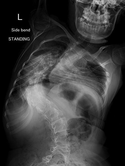
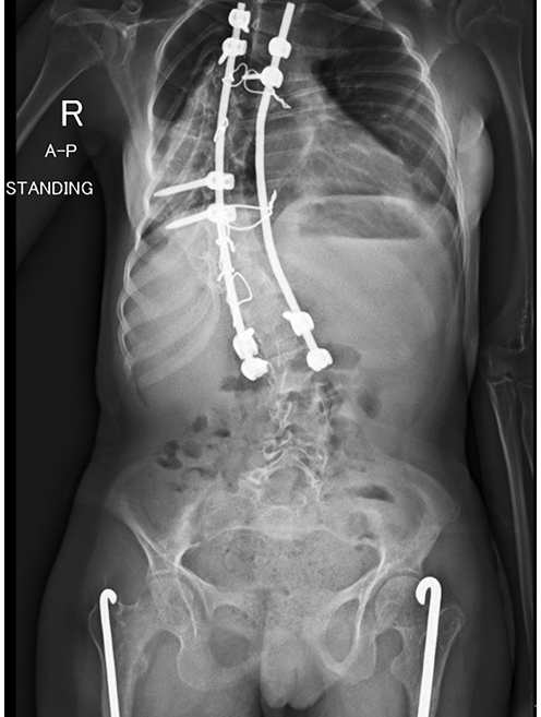
Figure 1. X-ray of patient’s spine before (on the left) and after (on the right) spine correction procedure.
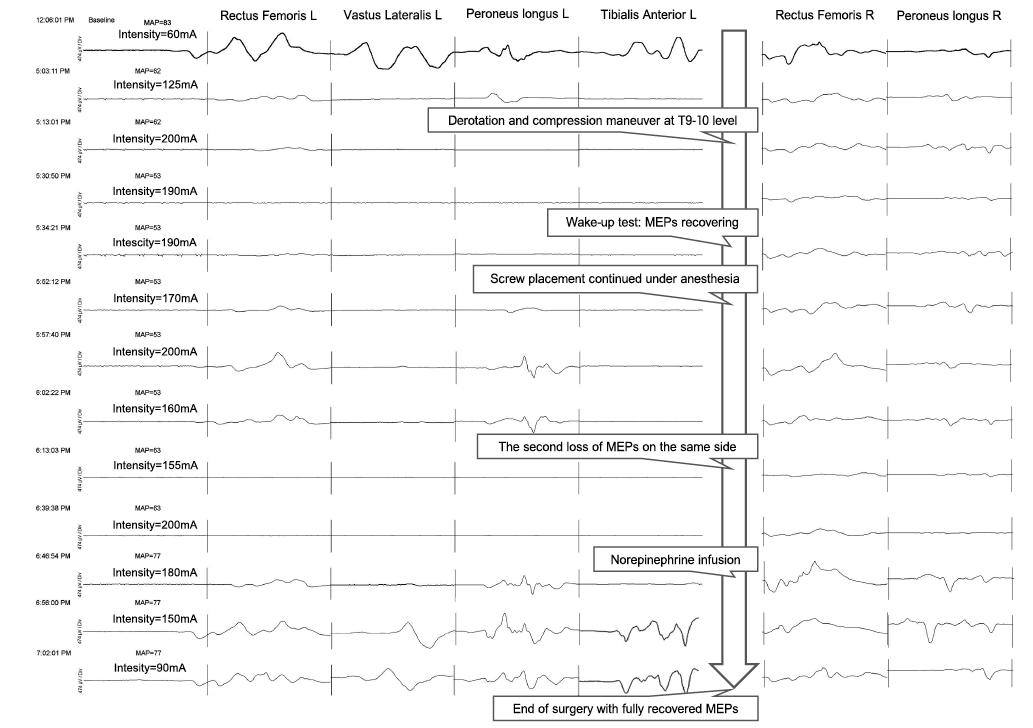
Figure 2. Motor evoked potential (MEP) curves obtained using Inomed ISIS Xpress IONM system (parameters: pulse width 1 ms, frequency 1.2 Hz) with NeuroExplorer 7 software. MAP – mean arterial pressure (mm Hg).
References
- Schwartz DM, Auerbach JD, Dormans JP et al. Neurophysiological detection of impending spinal cord injury during scoliosis surgery. J Bone Joint Surg Am. 2007;89(11):2440-9.
- Lewis SJ, Wong IHY, Strantzas S et al. Responding to intraoperative neuromonitoring changes during pediatric coronal spinal deformity surgery. Global Spine J. 2019;9(1 Suppl):15S-21S.
- Segura MJ, Talarico ME, Miranda MÁ, Noel MA. Analysis of intraoperative motor evoked potential changes and surgical interventions in 513 pediatric spine surgeries. J Clin Neurophysiol. 2024;41(2):161-8.
- Angelliaume A, Alhada TL, Parent HF, Royer J, Harper L. Intraoperative neurophysiological monitoring in scoliosis surgery: literature review of the last 10 years. Eur Spine J. 2023;32(9):3072-6.
- Ushirozako H, Yoshida G, Hasegawa T et al. Characteristics of false-positive alerts on transcranial motor evoked potential monitoring during pediatric scoliosis and adult spinal deformity surgery: an “anesthetic fade” phenomenon. J Neurosurg Spine. 2019;32(3):423-31.
- Miller SM, Donegan SW, Voigt N et al. Transcranial motor-evoked potentials for prediction of postoperative neurologic and motor deficit following surgery for thoracolumbar scoliosis. Orthop Rev (Pavia). 2019;11(1):7757.