


Sleep is a fundamental biological function. It is now recognized as one of the essential components of general well-being, as well as of health components, in particular, cardiometabolic and brain health. In 2021, American Heart Association included sleep duration as an essential component of cardiovascular health in the concept of Life’s essential 8 [1]. Nevertheless, sleep health is much beyond just sleep duration, it is a multidimensional concept which comprises several dimensions, consi-
ders adaptation to individual, social, and environmental demands that promotes physical and mental well-being [2]. This concept, initially suggested by D.J. Buysse [2], was largely developed in the last decade, and has recently been summarized by A. Voster et al. (2024) with the new 8 dimensions of sleep health: regularity, satisfaction, alertness/sleepiness, timing, efficiency, duration, breathing, and disordered sleep [3]. The impact of these dimensions on health can be independent or interrelated, enhan-
cing or modifying health risks and outcomes (Table) [3]. However, this approach does not consider some other sleep metrics which impact health. In particular, sleep architecture, sleep microstructure, sleep fragmentation, sleep stability, etc. although related to some of these 8 dimensions, are not integrated in the concept of multidimensional sleep pattern. This gap is partly filled in by the scientific statement on implications of sleep health on cardiometabolic health published by the American Heart Association on 14 April 2025 which included sleep architecture as one of sleep dimensions (Table) [4].
Although the evidence is rather limited, the alterations in sleep architecture were found in acute cardiocerebrovascular events and were shown to be associated with outcomes. A number of studies report significant changes in sleep architecture in post-stroke patients compared to control subjects [5]. According to a meta-analysis, these include a greater S1 percentage, a lower S2 percentage, and a reduced slow wave sleep (SWS) percentage and duration [6]. Some authors also report decreased rapid eye movement (REM) sleep percentage [7-9]. The impairment in sleep architecture is associated with the worse outcomes in stroke patients [10]. In particular, poor short-term outcomes were associated with reduced non-REM (NREM) stage 2 [11] and increased NREM stage 1 sleep [10], reduction of REM sleep together with a prolonged REM sleep latency was also linked with poor outcome (assessed by modified Rankin Scale) [7], and lower REM percentage was associated with the higher National Institutes of Health Stroke Scale
(NIHSS) at discharge [12] and poorer functional outcomes at 3 months (Barthel Index, modified Rankin Scale) [13]. It should be highlighted that there is a substantial individual variability in the follow-up changes in sleep architecture in post-stroke patients [11], which has also been reported from the longitudinal cohort studies [14].
Less evidence is available regarding sleep structure in acute cardiovascular events. Based on a systematic
review by M.T. Madsen et al. (2019), SWS, and REM sleep are reduced immediately following acute coronary syndrome (ACS) and significantly increase at 6-month follow-up [15]. These changes are associated with the increase in NREM stage 1 and 2. Patients with myocardial infarction (MI) also demonstrate higher sleep fragmentation. Patients with refractory angina have significantly reduced SWS compared to patients with stable angina. However, data regarding the association of sleep architecture changes and prognosis is lacking and limited to a brief report by the Japanese scientists (Nakashima et al., 2013) who showed that the decreased NREM stage 2 was an independent predictor of the recurrence of ACS and major adverse cardiovascular events (MACE) (hazard ratios: ACS, 2.17, p = 0.034; MACE, 1.87, p = 0.037) [16].
A Brazilian study demonstrated that maintenance of SWS is associated with longevity. On the other hand, inadequate sleep (measured by both subjective sleep quality
and objective sleep fragmentation) is associated with the accelerated age-related brain aging [17]. M. Scullin and C. Gao (2018) hypothesize that a dynamic interaction between SWS and REM sleep throughout life and aging plays role in cognitive health and cognitive longevity [14].
The need for objective evaluation by polysomnography results in rather scarce longitudinal evidence of the association of sleep structure changes and long-term outcomes [14,18]. It also explains the lack of these parameters in the early versions of Sleep Health concept.
The initial concept of sleep health developed by
D.J. Buysse (2014), implied self-reported (subjective) evaluation of its components, namely SATED (Satisfaction, Alertness, Timing, Efficiency, Duration) [2]. The later modifications consider both subjective and objective measurements [3,4]. Thus, some of the components of sleep health are estimated based on subjective assessments (e.g. satisfaction), some can be evaluated both subjectively and objectively (e.g. duration, efficiency, sleepiness, breathing), while others require objective confirmation (e.g. sleep structure) (Table).
Polysomnography (PSG) is the gold standard for objective sleep assessment, and implies recording of electroencephalogram (EEG), electro-oculogram, electromyogram from masseter muscles and limb muscles, respiration, thoracoabdominal movements, electrocardiogram (ECG) and other signals. It provides accurate data on sleep macro- and micro-structure, awakenings, sleep-related breathing disorders, limb movements and other movement disorders, etc. However, it is not the optimal strategy, as it provides a single night measure in artificial conditions (sleep lab) that might lead to sleep disturbance [19]. Moreover, it cannot define sleep timing, sleep rhythmicity and shows limited correlation with subjective metrics [20,21]. There is a substantial interrater
variability in sleep scoring, and it depends on various factors (patient characteristics [22], scorer training, etc. [23]). In addition, PSG is costly, time- and effort-consuming, and not reimbursed in many countries [24-26].
Nevertheless, the novel technologies and the develop-
ment of machine-learning methods, and in particular, deep learning techniques, are changing the field rapidly,
introducing alternative approaches to automatic sleep stage classification based on other signals rather than EEG [27]. Clear sleep-stage dependent fluctuations in the activity of autonomic nervous system [28] include an increase in parasympathetic tone during transition from wake to sleep and from light to deep sleep, and an increase in sympathetic activity with abrupt sympathetic surges during REM-sleep [29]. These changes are associated with the variations in heart rate variability (HRV), respiration, circulation, skin resistance, etc. providing opportunity to estimate sleep stages from ECG [30,31], respiratory signals, photoplethysmography (PPG) [32-34], electrodermal activity [35], temperature, movements [36], etc. or their combination [36-39]. The signals can be obtained from polygraphy/cardiorespiratory monitoring, pulse oximetry, actigraphy [40], wearables [32,41], and even non-contact monitoring systems [42-44]. In the majority of studies, the derived signals rather than raw data are used (e.g. heart rate, HRV rather than ECG, respiratory intervals rather than thoracoabdominal movements, etc.). The approach usually includes several steps: signal collection, extraction of derived time series (derived signals), preprocessing, and neural network classification [27,39].
Based on the data derived from the mentioned signals or their combination, different sleep-stage classifications can be developed: two-stage classification differentiating sleep and wakefulness [45,46], three-stage – NREM/REM/wakefulness, four-stage – light sleep/deep sleep/REM/wakefulness [30], and five-stage – N1/N2/N3/REM/wakefulness [34], with three- and four-stage classification being the most common and well-performed [27].
Sleep staging based on routine physiological signs is highly demanded in patients with cardiovascular diseases and acute events when the performance of full PSG is limited due to the patient severe condition, potential cognitive impairment, immobilization, conditions in the intensive care units (ICU), and other factors. However, the currently published algorithms were mainly developed and tested in healthy cohorts [32,33,37,38,47] or patients with sleep-disordered breathing [48] with the exclusion of patients with known cardiovascular and neurologic diseases. The latter ones, as well as patients with metabolic diseases, are often characterized by autonomic dysfunction and impaired HRV, which might limit the application of automated sleep stage classification. Moreover, a number of medications commonly used in cardiometabolic pathology affect autonomic nervous system. The results of L. Hermans et al. (2024) who assessed the performance of cardiorespiratory-based algorithm for sleep staging in patients using beta-blockers are promising [49]. They showed substantial agreement for four-class sleep staging (for heart rate and respiratory effort: kappa 0.635 (0.558; 0.700), accuracy 78.1 (74.5; 81.2) % versus kappa 0.660 (0.598; 0.736), accuracy 78.8 (74.8; 83.7) % for patients using beta-blockers and controls, respectively, p = 0.127; for ECG/heart rate only: kappa 0.535 (0.430; 0.632), accuracy 73.4 (65.4; 77.9) % versus kappa 0.535 (0.433; 0.629), accuracy 71.2 (66.8; 78.7) % for patients using beta-blockers and controls, respectively, p=0.127), and confirmed that despite potential impact of beta-blockers on HRV, it does not interfere with a reliable performance of the algorithm. At the same time, the studies evaluating reliability of algorithms in patients using other drugs affecting autonomic system are lacking [50,51]. The few studies that evaluated performance of automated sleep staging in patients with neurologic diseases [52] and different sleep disorders [53], demonstrated poorer classification performance [54] (in particular, for patients with REM parasomnia).
P.-W. Chen et al. (2022) evaluated the performance of automated sleep staging in 10 patients with stroke within inpatient rehabilitation unit. They used several signals obtained from wearable sensors, enabling recording of ECG, heart rate, respiratory rate, proximal skin temperature, and PPG for SpO2, peripheral arterial tonometry, and distal skin temperature [55]. They applied different 2 types of models (population and personal) and 4 algorithms (Balanced Bagging, Balanced Random Forest, Gradient Boosting, and XGBoost). According to their analysis, the performance of the
population models was rather poor (the highest Cohen’s kappa 0.27 by Gradient Boosting for 2stage sleep classification). Personalized models performed better (the highest Cohen’s kappa 0.66 for 2-stage classification, 0.60 for 3-stage, and 0.531 for 4-stage sleep classification for XGBoost algorithm). The low performance of the population models is likely to be related to the between-patient heterogeneity in physiological signals. Although personal model showed to be more accurate in stroke patients, it requires a PSG-reference for each patient [55].
In our preliminary study (unpublished data), in a small sample of stroke patients (n = 9) the performance of cardiorespiratory-based algorithm appeared to be highly variable with Cohen’s kappa achieving 0.79 (Figure). These data outperform published algorithms; however, a larger cohort is needed to verify these preliminary data.
Another gap in the field is the lack of algorithms for long-term sleep monitoring and sleep staging using non-EEG data which is partly explained by the substantial between- and within-subject variability of the assessed signals and the effects of confounding factors (age, gender, body mass index, time of the night, etc.) [56]. W. Ganglberger et al. (2023) assessed the validity of algorithms for sleep staging with long-term monitoring of HRV and respiratory signals in ICU patients [57]. The models showed lower agreement for ICU versus sleep laboratory data (60% versus 81%). However, they excluded patients with pre-existing cardiovascular and neurologic diseases and cognitive impairment.
To conclude, assessment of the components of sleep health in routine clinical practice is important. Sleep architecture, being one of the dimensions of sleep health, requires objective measurement by PSG that till nowadays has limited implementation. However, the rapid development of machine learning techniques opens the door to wider measurement of sleep parameters. Reliable and well-performing algorithms for sleep stage classification based on various physiologic parameters have been developed, although they are lacking for patients with cardiovascular diseases. Our preliminary data confirm that non-EEG-based sleep staging is feasible in stroke patients and good agreement can be achieved (kappa over 0.70). However, more studies are needed in this field. There exist some challenges decreasing agreement with the sleep scoring software preventing its immediate clinical implementation, including comorbid pathology, high between- and within-subject variability, confounders affecting physiologic measures (autonomic activity), etc. T. Penzel, an international expert in the field, suggests that novel methods are needed, for example, using the approach based on probabilities for sleep stages rather than single gold reference, and the development of a reference database for the validation of computer-based automated sleep scoring [58].
Conflict of interest
Authors declare no conflict of interest.
Funding
The work is supported by the grant of the Russian Scientific Foundation 25-25-00307.
Table. The multidimensional concept of sleep health (2014-2025) (adapted from [2-4]).
Parameter | Description | Buysse (2014) | Vorster et al. (2024) | AHA Statement, | Assessment | Health risks and outcomes |
Satisfaction/Quality | The subjective perception of the quality (“good” or “poor”) of sleep | + | + | + | Subjective (questionnaires, e.g. PSQI, sleep diary) | Mortality, MS, GMI, HTN, CHD, depression |
Alertness / Sleepiness / Napping | The ability to sustain optimal vigilance throughout waking hours | + | + | + | Subjective (ESS, KSS, sleep diary) Objective (MSLT, MWT, PVT, actigraphy – for napping) | Mortality, cardiovascular mortality, CHD, altered cognitive performance |
Timing | The placement of sleep within the 24-hour period | + | + | + | Subjective (questionnaire, sleep diary) Objective (actigraphy) | Mortality, CHD, MS, GMI, altered psychomotor performance |
Efficiency (or sleep continuity) | The ease of falling asleep and returning to sleep | + | + | + | Subjective (questionnaire, sleep diary) Objective (actigraphy, PSG) | Mortality, MS, GMI, HTN, CHD, depression, emotional distress, neurodegenerative diseases, altered psychomotor performance |
Duration | The total amount of sleep obtained per 24 hours | + | + | + | Subjective (questionnaire, sleep diary) Objective (actigraphy, PSG) | Mortality, obesity, MS, GMI, HTN, CHD, altered psychomotor performance |
Regularity/rhythmicity | Night-by-night regularity of bedtimes and get-up times, stability of sleep-wake patterns over time | - | + | + | Subjective (questionnaire, sleep diary) Objective (actigraphy) | Mortality, MS, GMI, depression |
Breathing (snoring) | Sleep-disordered breathing | - | + | Included in “sleep disorders” | Subjective (questionnaires) Objective (PSG, polygraphy) | CVD, mortality, altered cognitive performance, MS |
Disordered/disturbed sleep | The presence and severity of sleep disturbances | - | + | + | Subjective (questionnaires) Objective (PSG, polygraphy, actigraphy) | CVD, MS, neurodegenerative diseases |
Sleep architecture | Sleep staging (amount and temporal organization of sleep stages) | - | - | + | Objective (PSG) | ? |
CHD – coronary heart disease, CVD – cardiovascular disease, ESS – Epworth sleepiness scale, GMI – glucose metabolism impairment, HTN – hypertension, KSS – Karolinska sleepiness scale, MS – metabolic syndrome, MSLT – multiple sleep latency test, MWT – maintenance of wakefulness test, PSQI – Pittsburgh Sleep Quality Index, PSG – polysomnography, PVT – psychomotor vigilance test. | ||||||
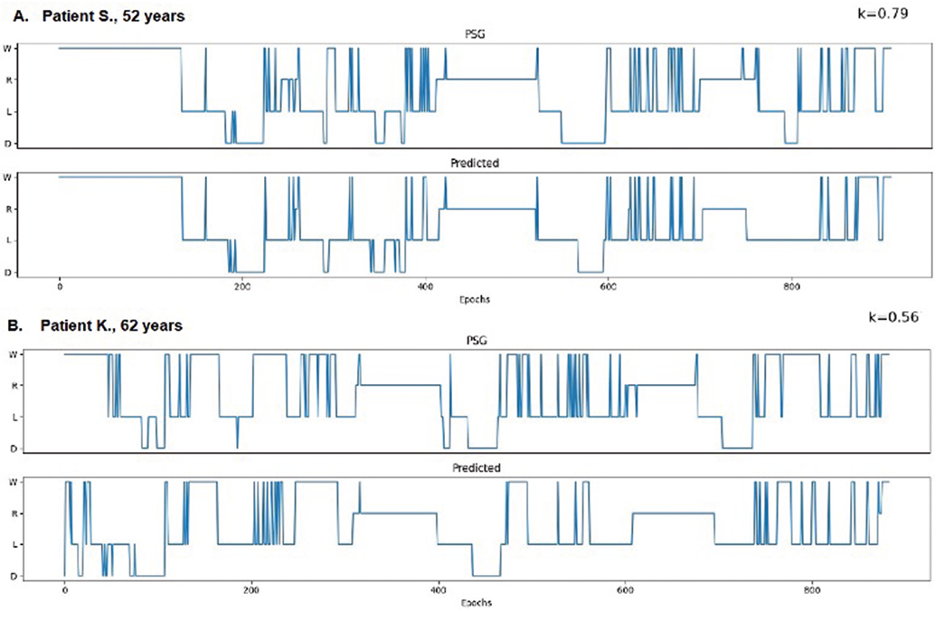
Figure. Hypnograms (scheme of sleep architecture) of patients with acute stroke based on manual scoring of polysomnogram and predicted by algorithm from sleep staging from cardiorespiratory parameters (unpublished data). A. Male patient with lacunar left middle cerebral artery ischemic stroke. NIHSS: at admission 7, at discharge 3; modified Rankin scale: at admission 2, at discharge 2. B. Male patient with left middle cerebral artery ischemic stroke of undetermined etiology. NIHSS: at admission 7, at discharge 2; modified Rankin scale: at admission 3, at discharge 3. Four-stage classification of sleep: W – wakefulness, R – REM-sleep, L – light sleep, D – deep sleep; NIHSS – National Institutes of Health Stroke Scale; PSG – polysomnography; predicted – sleep stages predicted by the developed algorithms based on cardiorespiratory data.
References
- Lloyd-Jones DM, Allen NB, Anderson CAM, et al. Life’s Essential 8: Updating and Enhancing the American Heart Association’s Construct of Cardiovascular Health: A Presidential Advisory from the American Heart Association. Circulation 2022;146:E18–43.
- Buysse DJ. Sleep Health: Can We Define It? Does It Matter? Sleep 2014;37:9–17.
- Vorster APA, van Someren EJW, Pack AI, et al. Sleep Health. Clinical and Translational Neuroscience 2024;8:8.
- St-Onge M-P, Aggarwal B, Fernandez-Mendoza J, et al. Multidimensional Sleep Health: Definitions and Implications for Cardiometabolic Health: A Scientific Statement From the American Heart Association. Circ Cardiovasc Qual Outcomes 2025;18:e000139.
- Ferre A, Ribó M, Rodríguez-Luna D, et al. Strokes and their relationship with sleep and sleep disorders. Neurologia 2013;28:103–18.
- Baglioni C, Nissen C, Schweinoch A, et al. Polysomnographic characteristics of sleep in stroke: A Systematic review and meta- analysis. PLoS One 2016;11:e0148496.
- Pace M, Camilo MR, Seiler A, et al. REM sleep as predictor of functional outcome after stroke: a translational study. Sleep 2018;41.
- Koerner E, Flooh E, Reinhart B, et al. Sleep alterations in ischemic stroke. Eur Neurol 1986;25:104–10.
- Kurushina OV, Kurakova EA. Sleep architecture of patients in the early recovery period after ischemic or hemorrhagic stroke. Clinical Review for General Practice 2023;4:14–8.
- Vock J, Achermann P, Bischof M, et al. Evolution of sleep and sleep EEG after hemispheric stroke. J Sleep Res 2002;11:331–8.
- Bassetti CL, Aldrich MS. Sleep electroencephalogram changes in acute hemispheric stroke. Sleep Med 2001;2:185–94.
- Miano S, Fanfulla F, Nobili L, et al. SAS CARE 1: Sleep architecture changes in a cohort of patients with Ischemic Stroke/TIA. Sleep Med 2022;98:106–13.
- Denis C, Jaussent I, Guiraud L, et al. Functional recovery after ischemic stroke: Impact of different sleep health parameters. J Sleep Res 2024;33:e13964.
- Scullin MK, Gao C. Dynamic Contributions of Slow Wave Sleep and REM Sleep to Cognitive Longevity. Curr Sleep Med Rep 2018;4:284–93.
- Madsen MT, Huang C, Zangger G, et al. Sleep disturbances in patients with coronary heart disease: A systematic review. Journal of Clinical Sleep Medicine 2019;15:489–504.
- Nakashima H, Henmi T, Minami K, et al. Impact of sleep structure on long-term clinical outcomes in patients with acute myocardial infarction . Eur Heart J, 2013, p. P1323.
- Ramduny J, Bastiani M, Huedepohl R, et al. The association between inadequate sleep and accelerated brain ageing. Neurobiol Aging 2022;114:1–14.
- Ohayon MM, Carskadon MA, Guilleminault Ch, et al. Meta-Analysis of Quantitative Sleep Parameters From Childhood to Old Age in Healthy Individuals: Developing Normative Sleep Values Across the Human Lifespan. Sleep 2004;27:1255–73.
- Bruyneel M, Sanida C, Art G, et al. Sleep efficiency during sleep studies: Results of a prospective study comparing home-based and in-hospital polysomnography. J Sleep Res 2011;20:201–6.
- Allen SF, Elder GJ, Longstaff LF, et al. Exploration of potential objective and subjective daily indicators of sleep health in normal sleepers. Nat Sci Sleep 2018;10:303–12.
- Danker-Hopfe H, Kunz D, Gruber G, et al. Interrater reliability between scorers from eight European sleep laboratories in subjects with different sleep disorders. J Sleep Res 2004;13:63–9.
- Zhang X, Dong X, Kantelhardt JW, et al. Process and outcome for international reliability in sleep scoring. Sleep and Breathing 2015;19:191–5.
- Magalang UJ, Chen NH, Cistulli PA, et al. Agreement in the scoring of respiratory events and sleep among international sleep centers. Sleep 2013;36:591–6.
- Khachatryan SG, Isayan MA, Hovakimyan HA. Current state and future perspectives of sleep medicine and sleep research in Armenia. A Joint Position Statement of Armenian Sleep Disorders Association, Armenian National Institute of Health, and European Assembly of National Sleep Societies. Armenian Journal of Health & Medical Sciences 2021;1:28–40.
- Parish JM, Freedman NS, Manaker S. Evolution in reimbursement for sleep studies and sleep centers. Chest 2015;147:600–6.
- Gjerstad MD, Puertas J, Goncalves M, et al. Reimbursement of Sleep Procedures across Europe. 2014.
- Liu P, Qian W, Zhang H, et al. Automatic sleep stage classification using deep learning: signals, data representation, and neural networks. Artif Intell Rev 2024;57:301.
- Tobaldini E, Nobili L, Strada S, et al. Heart rate variability in normal and pathological sleep. Front Physiol 2013;4:1–11.
- Shinar Z, Akselrod S, Dagan Y, et al. Autonomic changes during wake-sleep transition: A heart rate variability based approach. Auton Neurosci 2006;130:17–27.
- Li Q, Li Q, Liu C, et al. Deep learning in the cross-time frequency domain for sleep staging from a single-lead electrocardiogram. Physiol Meas 2018;39:124005.
- Kesper K, Canisius S, Penzel T, et al. ECG signal analysis for the assessment of sleep-disordered breathing and sleep pattern. Med Biol Eng Comput 2012;50:135–44.
- Walch O, Huang Y, Forger D, et al. Sleep stage prediction with raw acceleration and photoplethysmography heart rate data derived from a consumer wearable device. Sleep J 2019;42.
- Ye Y, Yang K, Jiang J, et al. Automatic Sleep and Wake Classifier with Heart Rate and Pulse Oximetry: Derived Dynamic Time Warping Features and Logistic Model. 2016 Annual IEEE Systems Conference, IEEE; 2016, p. 1022.
- Korkalainen H, Aakko J, Duce B, et al. Deep learning enables sleep staging from photoplethysmogram for patients with suspected sleep apnea. Sleep J 2020;43.
- Herlan A, Ottenbacher J, Schneider J, et al. Electrodermal activity patterns in sleep stages and their utility for sleep versus wake classification. J Sleep Res 2018;28:e12694.
- Gaiduk M, Perea JJ, Seepold R, et al. Estimation of Sleep Stages Analyzing Respiratory and Movement Signals. IEEE J Biomed Health Inform 2022;26:505–14.
- Redmond SJ, De Chazal P, O’Brien C, et al. Sleep staging using cardiorespiratory signals. Somnologie 2007;11:245–56.
- Beattie Z, Oyang Y, Statan A, et al. Estimation of sleep stages in a healthy adult population from optical plethysmography and accelerometer signals. Physiol Meas 2017;38:1968–79.
- Tataraidze A, Korostovtseva L, Anishchenko L, et al. Sleep architecture measurement based on cardiorespiratory parameters. Proceedings of the Annual International Conference of the IEEE Engineering in Medicine and Biology Society, EMBS, 2016.
- Yuan H, Hill EA, Kyle SD, et al. A systematic review of the performance of actigraphy in measuring sleep stages. J Sleep Res 2024;33:e14143.
- Svensson T, Chung U il, Tokuno S, et al. A validation study of a consumer wearable sleep tracker compared to a portable EEG system in naturalistic conditions. J Psychosom Res 2019;126:109822.
- Wang Z, Zha S, Yu B, et al. Sleep Staging Using Noncontact-Measured Vital Signs. J Healthc Eng 2022;2022:2016598.
- Tataraidze A, Korostovtseva L, Anishchenko L, et al. Bioradiolocation-based sleep stage classification. Annu Int Conf IEEE Eng Med Biol Soc, 2016, p. 2839–42.
- Tataraidze A, Anishchenko L, Korostovtseva L, et al. Sleep stage classification based on bioradiolocation signals. Annu Int Conf IEEE Eng Med Biol Soc, 2015, p. 362–5.
- Casal R, Di Persia LE, Schlotthauer G. Classifying sleep–wake stages through recurrent neural networks using pulse oximetry signals. Biomed Signal Process Control 2021;63:102195.
- Casal R, Di Persia LE, Schlotthauer G. Sleep-wake stages classification using heart rate signals from pulse oximetry. Heliyon 2019;5:e02529.
- Cerina L, Overeem S, Papini GB, et al. A sleep stage estimation algorithm based on cardiorespiratory signals derived from a suprasternal pressure sensor. J Sleep Res 2024;33:e14015.
- Abdul Motin M, Kamakar C, Marimuthu P, et al. Photoplethysmographic-based automated sleep-wake classification using a support vector machine. Physiol Meas 2020;41:075013.
- Hermans L, Meulen F van, Anderer P, et al. Performance of cardiorespiratory-based sleep staging in patients using beta blockers. Journal of Clinical Sleep Medicine 2024;20:575–81.
- Fiani D, Campbell H, Solmi M, et al. Impact of antidepressant use on the autonomic nervous system: A meta-analysis and systematic review . Eur Neuropsychopharmacol 2023:75–95.
- Krishnaiyan R, Thompson JP. Drugs affecting the autonomic nervous system. Anaesthesia and Intensive Care Medicine 2013;14:548–53.
- Macea J, Heremans ERM, Proost R, et al. Automated Sleep Staging in Epilepsy Using Deep Learning on Standard Electroencephalogram and Wearable Data. J Sleep Res 2025:e70061.
- Fonseca P, Van Gilst MM, Radha M, et al. Automatic sleep staging using heart rate variability, body movements, and recurrent neural networks in a sleep disordered population. Sleep 2020;43:1–10.
- van der Aar JF, van Gilst MM, van den Ende DA, et al. Hypnogram and Hypnodensity Analysis of REM Sleep Behaviour Disorder Using Both EEG and HRV-Based Sleep Staging Models. J Sleep Res 2025:e70046.
- Chen PW, O’Brien MK, Horin AP, et al. Sleep Monitoring during Acute Stroke Rehabilitation: Toward Automated Measurement Using Multimodal Wireless Sensors. Sensors 2022;22:1–15.
- Long X, Haakma R, Leufkens TRM, et al. Effects of between- and within-subject variability on autonomic cardiorespiratory activity during sleep and their limitations on sleep staging: A multilevel analysis. Comput Intell Neurosci 2015;2015:583620.
- Ganglberger W, Krishnamurthy PV, Quadri SA, et al. Sleep staging in the ICU with heart rate variability and breathing signals. An exploratory cross-sectional study using deep neural networks. Frontiers in Network Physiology 2023;3:1120390.
- Penzel T. Sleep scoring moving from visual scoring towards automated scoring. Sleep 2022;45:1–3.